The fact that dental X-rays involve exposure to radiation concerns some people. In reality, the amount of radiation used for a traditional X-ray is very small, especially when compared to the amount humans are exposed to in day-to-day life. The American Dental Association (ADA) and the Food and Drug Administration (FDA) have determined that they are safe for everyone, including children and pregnant women.
Radiation can, however, accumulate in the body over time. Dentists recognized the importance of limiting the number of X-rays so that build-up is kept to a minimum. Unless there is a good diagnostic reason, most dentists will only order X-rays once every 12 or 24 months. If a patient has a medical condition requiring numerous X-rays, the dentist may choose to forgo dental X-rays for that individual unless absolutely necessary.
Following Safety Guidelines
While there is little danger from all types of dental X-rays, the ADA and the FDA have collaborated to put guidelines in place to limit the risks even more for both patients and dental technicians. State and local health agencies may have their own guidelines as well.
Dental offices and their X-ray equipment are subject to state and local inspections and licensing. They adhere to certain procedures when doing X-rays. For example, lead aprons or capes are placed on the patient’s body to avoid exposure. Also, at one time, patients would hold the X-ray film in place themselves. Today, holders are used for this purpose. And technicians will step out of the room when X-rays are used to limit their own exposure.
Safer Technology
As technology has advanced, every type of dental X-ray has become even safer. Conventional X-rays use photographic film that is developed using chemicals in a dark room, much like photos are. Over time, improvements have been made to the film that is used in the process, so that patients are exposed to radiation for a shorter time.
Also, many dental offices are now using digital images instead of film. Not only is time saved in the development process, reducing the amount of radiation by as much as 80%.
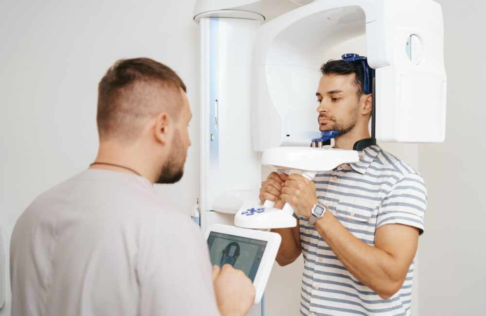
3D Imaging Using CBCT
An offshoot of dental X-rays is 3D imaging. Orthodontists and dentists use Cone Beam Computerized Tomography (CBCT), which evolved from CAT (Computerized Axial Tomography) Scan technology. More commonly known as Cone Beam Imaging, it creates a panoramic 3D image of a patient’s entire dental anatomy.
The scan time for a CBCT machine is very short, keeping a patient’s radiation exposure to a minimum. The technology provides images that are more accurate and detailed than 2D images. It gives a much more comprehensive view that shows the teeth, jaw, and joints, and how they all fit and move together in the skull.
X-Rays: The Best Diagnostic Tool
The benefits of getting an X-ray far outweigh the risks. With these types of dental X-rays, dentists have the ability to literally see what’s going on inside their patient’s teeth. They are able to diagnose issues like cavities early and fix them quickly, helping patients avoid unnecessary pain, cost, and further damage.