Dental X-rays: Types, Purpose, Procedure, and Risks

Dental X-rays can help your dentist detect oral health issues, like cavities and gum disease before they worsen. There are many different types of dental X-rays, including intraoral (taken inside your mouth) and extraoral (taken outside your mouth). Dental X-rays are essential to proper oral health and maintenance.
What are dental X-rays?
Dental X-rays (radiographs) are internal images of your teeth and jaws. Dentists use X-rays to examine structures they can’t see during a routine checkup, like your jawbone, nerves, sinuses, and teeth roots.
How do dental X-rays work?
Like X-rays taken in other parts of your body, dental X-rays use electromagnetic radiation to capture images of your mouth. The radiation beam passes through your soft tissues and creates images of your teeth and bones.
What can dental X-rays detect?

Dental X-rays help your dentist diagnose a wide range of oral health issues.
Dental X-rays show:
- Cavities, especially small areas of decay between teeth.
- Decay beneath existing fillings.
- Bone loss in your jaw
- Areas of infection.
- The position of unerupted or impacted teeth.
- Abscessed teeth (infection at the root of your tooth or between your gums and your tooth).
- Cysts and some types of tumors.
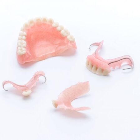
- Dentists also use X-rays to help determine your eligibility for treatments like dental implants, braces, or dentures. X-rays help your dentist check healing after certain procedures, too, such as dental bone grafts and root canal therapy.
How are dental X-rays done?
Before taking dental X-rays, a technician will place a lead apron over your chest and may wrap a thyroid collar around your neck. This helps protect you from excess radiation.
When it’s time to take the X-rays, you’ll either sit in a chair or stand in front of an X-ray machine. A technician will place the film or sensor, and then press a button to take the X-ray image. It’s important to hold as still as possible during this process.
What are the different types of dental X-rays?
There are two main types of dental X-rays:
- Intraoral: The film or sensor is inside your mouth.
- Extraoral: The film or sensor is outside your mouth.
Intraoral X-rays
There are different types of intraoral X-rays:
Bitewing X-rays
Bitewings show the upper and lower teeth in one area of your mouth. These dental X-rays help your dentist detect decay between your teeth or any changes that occur just below your gum line.
Bitewing X-rays don’t usually show the roots of your teeth.
Periapical X-rays
A periapical X-ray shows your entire tooth, from the crown to the root tip. This type of X-ray helps your dentist detect decay, gum disease, bone loss and any other abnormalities of your tooth or surrounding bone.
Occlusal X-rays
Occlusal X-rays help your dentist detect any issues in the floor or roof of your mouth. These images are helpful when diagnosing fractured or impacted teeth or evaluating the roots of your front teeth. Occlusal images can also help identify cysts, abscesses, and jaw fractures. Pediatric dentists may use occlusal X-rays to evaluate developing teeth.
Extraoral X-rays
There are several types of extraoral X-rays:
Panoramic X-rays
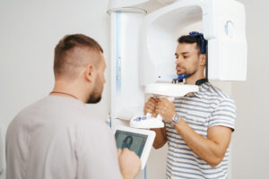
A panoramic dental X-ray shows all of the structures in your mouth on a single image, including your upper and lower teeth, jaw joints, nerves, sinuses, and supporting bone.
A panoramic X-ray allows your dentist to get an overview of any existing oral health issues.
Cephalometric X-rays
A cephalometric X-ray shows your entire head from the side. It shows your dentist the location of your teeth in relation to your jaw.
Orthodontists (dentists who specialize in correcting bites) often use cephalometric X-rays to plan treatment.
Cone beam CT scan
Dentists use a computed tomography (CT) scans to capture 3D dental X-rays of your teeth, jaws, joints, nerves and sinuses. These X-rays can also detect tumors or facial fractures.
Surgeons often use dental CT scans to check the height, width, and location of your jawbone before dental implant placement.
Are dental X-rays safe?
The radiation risk from a dental X-ray is quite small. In fact, the amount of radiation you get from a full set of dental X-rays is comparable to the amount of radiation you absorb from things like:
- TVs, smartphones, and computers.
- Building materials like ceramic floor tiles and granite countertops.
- Background radiation from the sun, stars, and the Earth itself.
In extremely large doses, however, dental X-rays can be harmful and may even increase your cancer risk. That’s why you shouldn’t have X-rays more often than necessary. Your healthcare provider can help you weigh the risks vs. benefits of dental X-rays.
How often should I get dental X-rays?
Most people with healthy teeth and gums should have dental X-rays taken once every six to 18 months. But if you have gum disease, recurring decay or other time-sensitive oral health issues, you may need more frequent X-rays.
Can I refuse dental X-rays?
As an individual, you have the right to refuse dental X-rays. However, it’s important to understand that most dentists won’t provide services without them.
If you’re concerned about radiation exposure, talk with your dentist. They can help you weigh the pros and cons of getting dental X-rays.
Can a dental X-ray show cancer?
Dental X-rays can show some types of oral cancer — particularly cancer that either started in or spread to your jaw. But X-rays can’t detect all types of mouth cancer. That’s why routine oral cancer screenings are so important.
Should I have dental X-rays while pregnant?
Generally speaking, it’s safe to have X-rays of your teeth while pregnant or breastfeeding (chestfeeding). In fact, both the American Dental Association and the American Pregnancy Association have stated that dental X-rays pose little to no risk to a fetus. Even so, most dentists avoid taking X-rays during pregnancy unless it’s absolutely necessary


